Find Your Focus: A 3D Guide to Choosing the Perfect Microscope
From the bustling pharma labs of Hyderabad to the cutting-edge research facilities in Bengaluru and quality control units in manufacturing plant of Pune, it is our window into the broad spectrum of life that would otherwise be invisible to the naked eye. But could be interested to know how these amazing instruments work. Or why are there so many different types of microscopes? At SIPCON Technologies, we affirm that tool knowledge drives great discovery.
Different Types of Microscopes
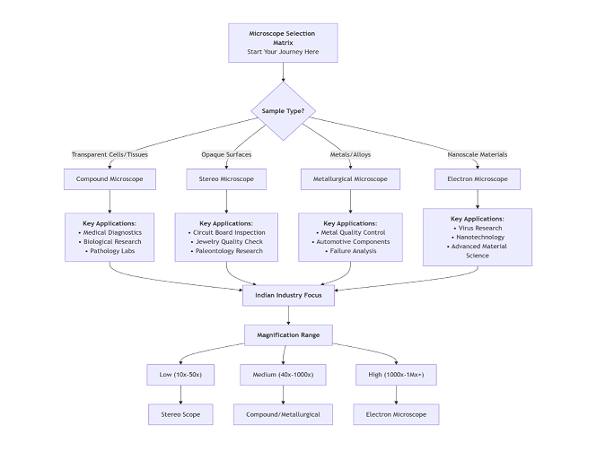
Compound Microscope, Our Old Friend: The workhorse of microscopy
This is what probably comes to mind when you think of the word “microscope.” It is a workhorse in schools, medical labs and for everyday biology.
Principle: It is based upon the principle of lens systems. That is done with the main lens (which is closer to the specimen) and the eyepiece lens (which is closer to your eye). The image of the specimen is provided in the microscope tube magnified real image by the main lens The eyepiece lens magnifies this key image again, providing your eye with a large virtual image.
Structure: Eyepiece: The lens through which you look.
Objective Turret: Rotating nosepiece like 4x, 10x, 40x, 100x are attached.
Stage: The place where you set the specimen slide.
Illuminator: The illumination located at the light-guide plate a condenser, directing it to the specimen.
Best for Use With: Thin transparent specimens like blood smears, bacteria, and cells.
3D Inspector – The Stereo Microscope
This is the same tool you will find on the benches of watchmakers and palaeontologists electronics quality control inspectors.
Principle: It is a low-power stereoscopic system. Opposite to the function of biological microscope, it employs two independent optical pathways (eyepieces and goals) that give your left and right eyes two slightly different angles of view. Your brain combines these, and you have 3D images of the specimen.
Structure: Two Eyepieces: It can be used for binocular and 3D viewing.
Two Independent Oculars: Each with a dedicated optical path
Top-Light Light source is reflected beam from above to use darkfield illumination.
Long Working Distance: The distance from the main lens to the object is longer for free movement of manipulating tools.
Best For: Looking at the surface of objects like circuit boards, fossils or insects while doing fine dissection.
Metallurgical Microscope: The Metalloid graphist
This device is useful for the microstructural study of materials and alloys in the automobiles and aeronautics industries
Principle – It is an incident beam microscope. the beam is used to form the image passes through the
All-Objective lens. Metals are dark. they are non- transmissive. The polished metal specimen is illuminated and does not need a reference beam. Once the beam reflects, it returns through the eyepiece, and will show the grain boundaries, phases, and inclusions.
Special Objectives: These are “epi-objectives” and serve dual purpose that is condenser and image lens.
Polarizing Filters – Included for polarization is PVC Hasteners. This is for polarization in material study.
Ideal Applications: Looking at the microstructures of metals, ceramics, and composites with respect to QC, heat treatment, and failure analysis.
Inverted Microscopy: A Friend to the Cell Cultivator
Walk into biology or biotechnology lab and chances are good you will see it.
Principle: It uses an inverted Optical design. The illuminator and condenser are located above the stage, and the main lenses below it. This lets users look up from the bottom of a specimen vessel like a cell culture flask or a Petri dish.
Structure: Ceiling Light: The source of illumination comes from above.
Objectives Under Object: Located under the stage.
Large Fixed Stage: Easily accommodates large culture dishes.
Ideal For: Viewing live cells that have been cultured in the bottom of a culture flask in liquid.
Electron Microscope: On the Trail of Atoms
In insufficient lighting the smallest of details, we rely on electrons. These are the engine rooms of nanotechnology & materials science.
Principle: Uses an accelerated beam of Electrons to image instead of photons (Light). because the electron wavelength is much shorter, it can have a vastly higher resolution. Magnet
Electron lenses for focusing the electron beam. There are 2 major types of this class of microscope:
Scanning Electron Microscope: A microscope that projects an intensified beam of electrons onto a specimen and produces its electron image signal It gives beautiful 3D like topographical images.
TEM (Transmission Electron Microscope): Sends a beam of electrons across a thin specimen. The image created by the electrons when they drift by. It can achieve atomic-level resolution.
Construction: Very sophisticated, contains an electron gun along with special electromagnetic lenses, a vacuum system, and dedicated detectors.
Best Uses: Observing viruses, nanoparticles, internal cell formation.

Conclusion and CTA
The Proper Window to the Micro World
Each of these microscope types is a work of optical engineering, created to address a particular challenge. It all depends on what you want to do with it: are you studying living cells? A microchip? Metal fatigue? Learning the principle and formation of them is the key to unleashing their full potential bringing your research or QC capabilities to new levels of accuracy. Ready to find the perfect microscope for you? It can be a little tricky trying to navigate the different types of microscopes alone though. At SIPCON Technologies, our experts are here to help.
Call us today for a free consultation. Let us assist you in choosing the perfect microscope for your job and budget so that you do not miss a single detail. We would love to connect at info@sipconinstrument.com, or check out our website https://www.sipconinstrument.com
Phone: +91 82229 29966


